Qing Dai
PhD Candidate at UCLA Bioengineering & Radiology

I’m a PhD candidate in Bioengineering at UCLA, working in the Magnetic Resonance Research Lab (MRRL), Department of Radiological Sciences, David Geffen School of Medicine, advised by Dr. Holden Wu. I’m also affiliated with the Jonsson Comprehensive Cancer Center.
I’m graduating in August 2026 and am actively looking for full-time roles in medical devices, surgical robotics, and imaging R&D — let’s connect!
Previously, I earned an M.S. in Biomedical Imaging at UCSF (advised by Dr. Peder Larson) and a B.S. in Biochemistry with a Bioinformatics minor at UCLA.
Focus Areas
- MRI-Guided Interventions
- Medical AI & Computer Vision
- Medical Device Integration
- Computational Modeling
Research
My research focuses on making minimally invasive interventions safer and more precise — building the imaging, AI, and computational tools that help clinicians see clearly and act precisely, right where it matters most.
My work spans two parts. (1) Technical development: real-time reconstruction and processing algorithms, machine-learning models for guidance and analysis, computational modeling, and integrated hardware/software systems — including two patent-pending technologies. (2) Full-arc validation: validating each method from phantom to ex vivo to in vivo across multiple imaging platforms and therapeutic modalities (microwave ablation, HIFU, biopsy, robotics), with clinical, engineering, and industry partners.
Updates
| Jun 02, 2026 | Our first-authored paper “Active Electromagnetic Interference Suppression for MR Thermometry During MR-Guided Microwave Ablation” was published in Magnetic Resonance in Medicine. Read more |
|---|---|
| May 12, 2026 | Received a Magna Cum Laude Merit Award at ISMRM 2026 (Cape Town) for our computational modeling abstract on pulsed microwave ablation. |
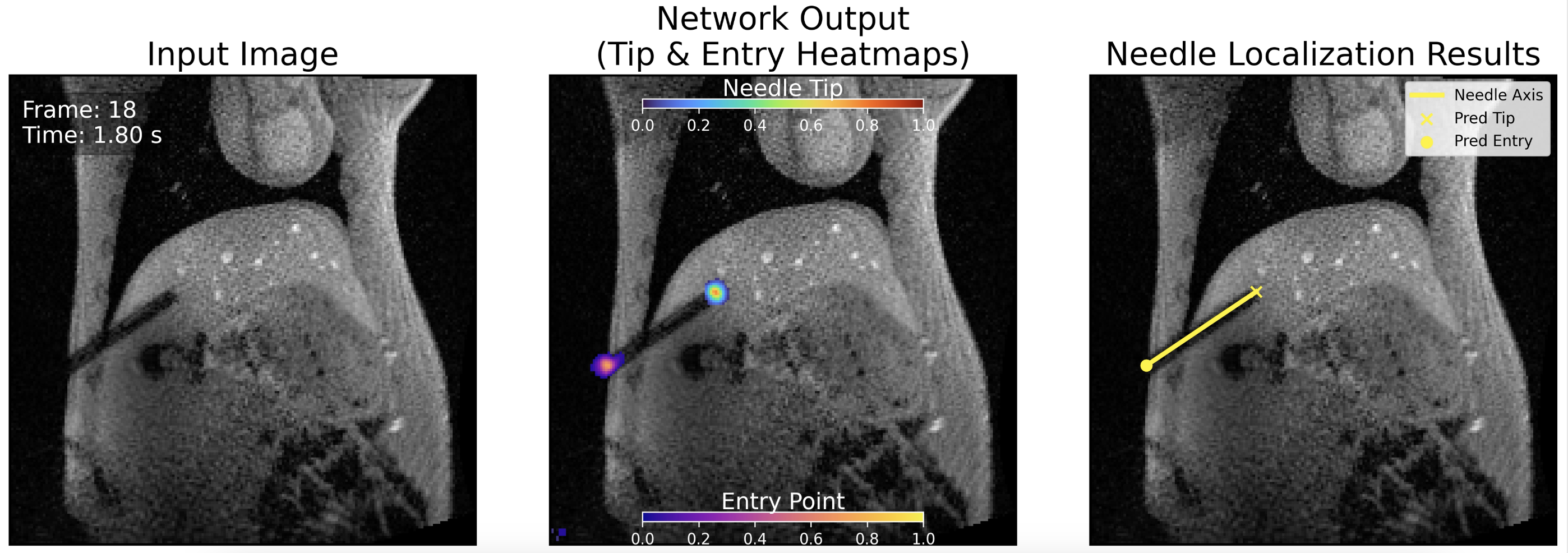
| Mar 30, 2026 | Our co-authored paper “Keypoint Detection Network for Needle Localization on Intra-Procedural MRI in MRI-Guided Liver Interventions” was published in IJCARS. Read more |
| Nov 17, 2025 | Our co-first-authored paper “Thermoresponsive Polymer-Modified SiO₂/Gadolinium-Diethylenetriamine Pentaacetic Acid Composite Nanoparticles for Magnetic Resonance Imaging-Guided Ultrasound-Modulated Contrast Enhancement at Human Body Temperatures” was published in ACS Applied Nano Materials. Read more |
| Sep 11, 2025 | Our first-authored paper “Volumetric Thermometry in Moving Tissues Using Stack-of-Radial MRI and an Image-Navigated Multi-Baseline Proton Resonance Frequency Shift Method” was published in Magnetic Resonance in Medicine. Read more |
Selected Publications
- iMRI
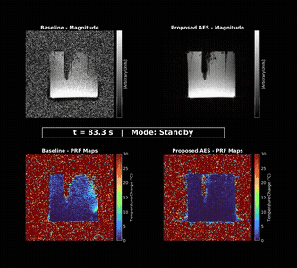 Active electromagnetic interference suppression for MR thermometry during MR-guided microwave ablationMagnetic Resonance in Medicine, 2026U.S. Patent Pending
 - iMRI
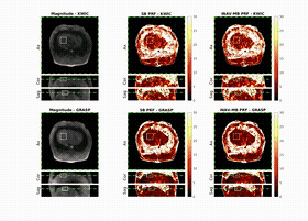 Volumetric thermometry in moving tissues using stack-of-radial MRI and an image-navigated multi-baseline proton resonance frequency shift methodMagnetic Resonance in Medicine, 2026