Research
My research focuses on making minimally invasive interventions safer and more precise — building the imaging, AI, and computational tools that help clinicians see clearly and act precisely, right where it matters most.
My work spans two parts. (1) Technical development: real-time imaging and reconstruction, machine-learning models for guidance and analysis, computational modeling, and integrated hardware/software systems — including two patent-pending technologies. (2) Full-arc validation: testing each method from phantom to ex vivo to in vivo across multiple imaging platforms and therapeutic modalities (microwave ablation, HIFU, biopsy, robotics), with clinical, engineering, and industry partners.
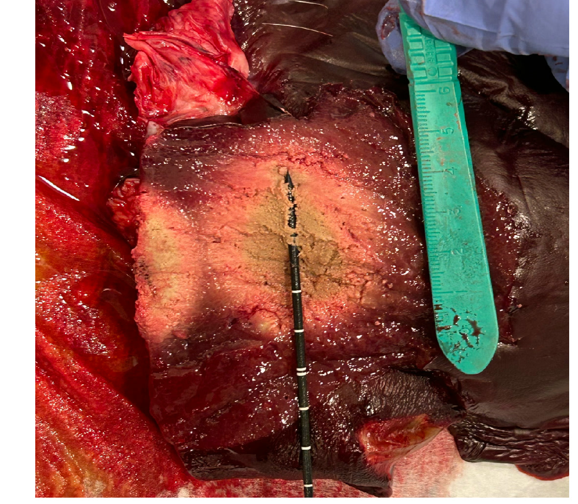
Real-time imaging and monitoring for MRI-guided thermal ablation
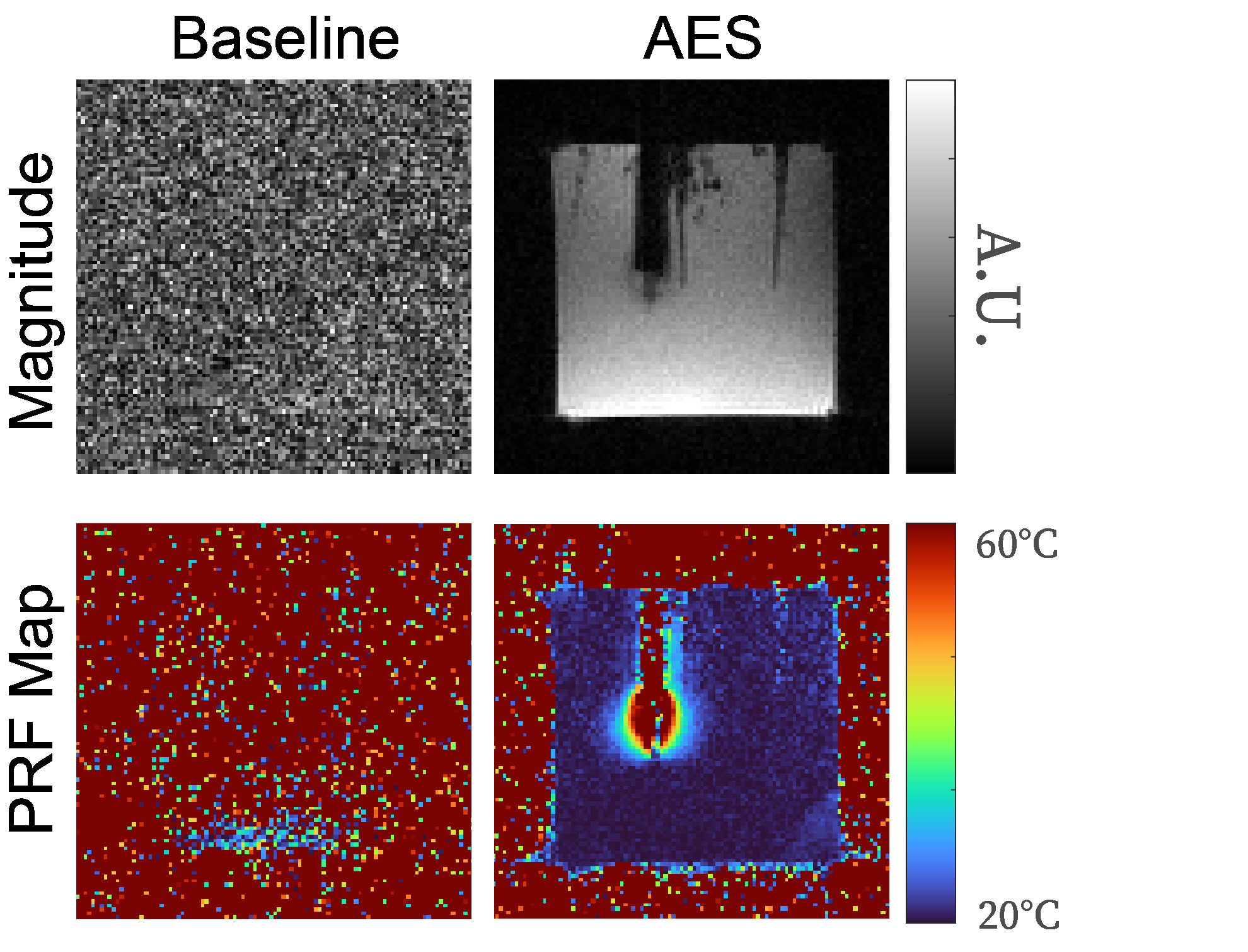
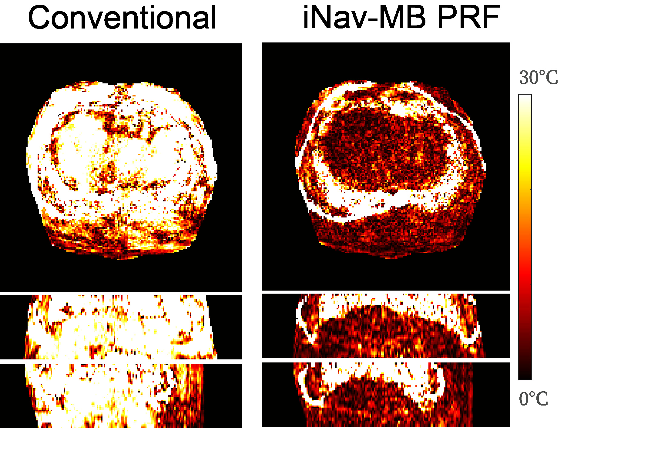
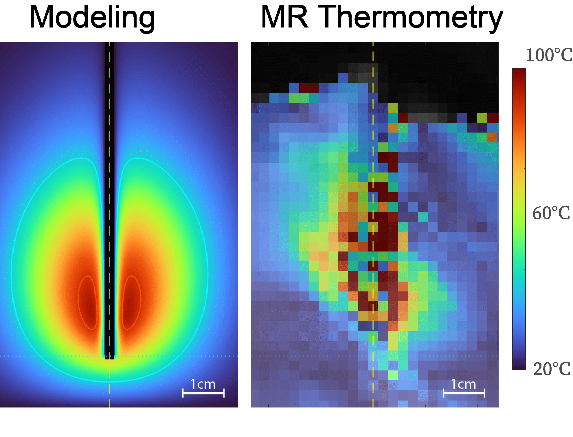
I develop imaging and computational methods that improve MRI-guided thermal ablation — including a software-based EMI suppression system that eliminates device interference without hardware modifications (patent pending) [1], a volumetric thermometry framework that tracks temperature across the entire liver during free-breathing with <1.5°C accuracy [2], and calibrated computational models that predict ablation outcomes for pre-procedural planning [3].
Signal ProcessingMotion TrackingImage ReconstructionComputational Modeling (EM & Bioheat Transfer)Real-Time Imaging
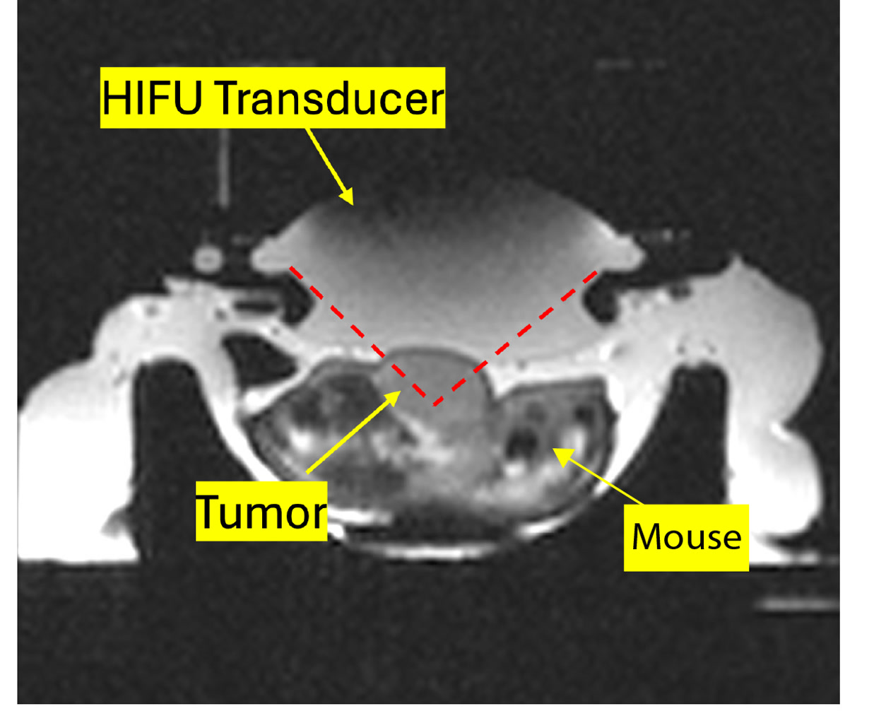
Theranostic imaging for nanoparticle drug delivery
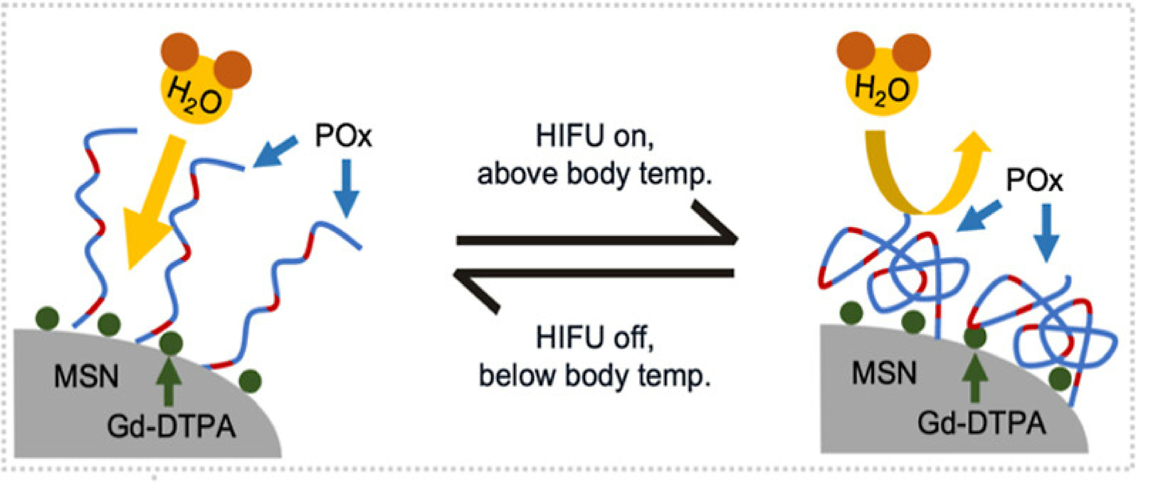
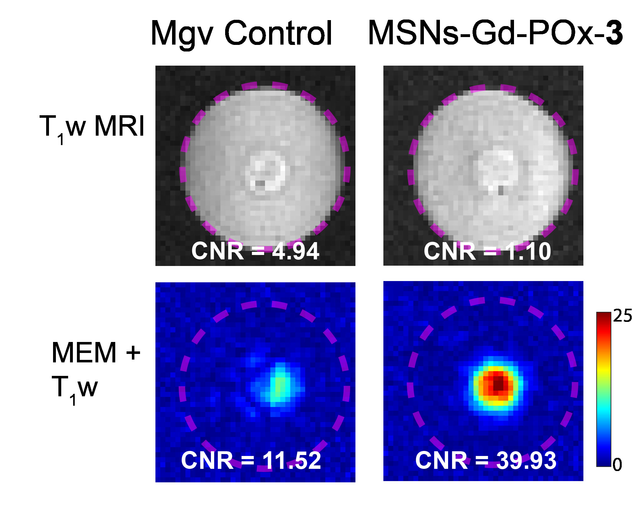
I develop MRI-based methods for confirming and monitoring nanoparticle-mediated drug delivery triggered by focused ultrasound [6]. This spans imaging pipeline design, HIFU protocol optimization, and in vivo validation — demonstrating spatially targeted visualization of nanoparticle delivery with 139× signal amplification and improved therapeutic outcomes in murine cancer models. In collaboration with the Zink Group (UCLA Chemistry & Biochemistry).
Contrast EnhancementHIFUNanoparticle Drug DeliveryTheranostic ImagingImaging Pipeline DesignIn Vivo Validation
Machine learning and computer vision for medical imaging
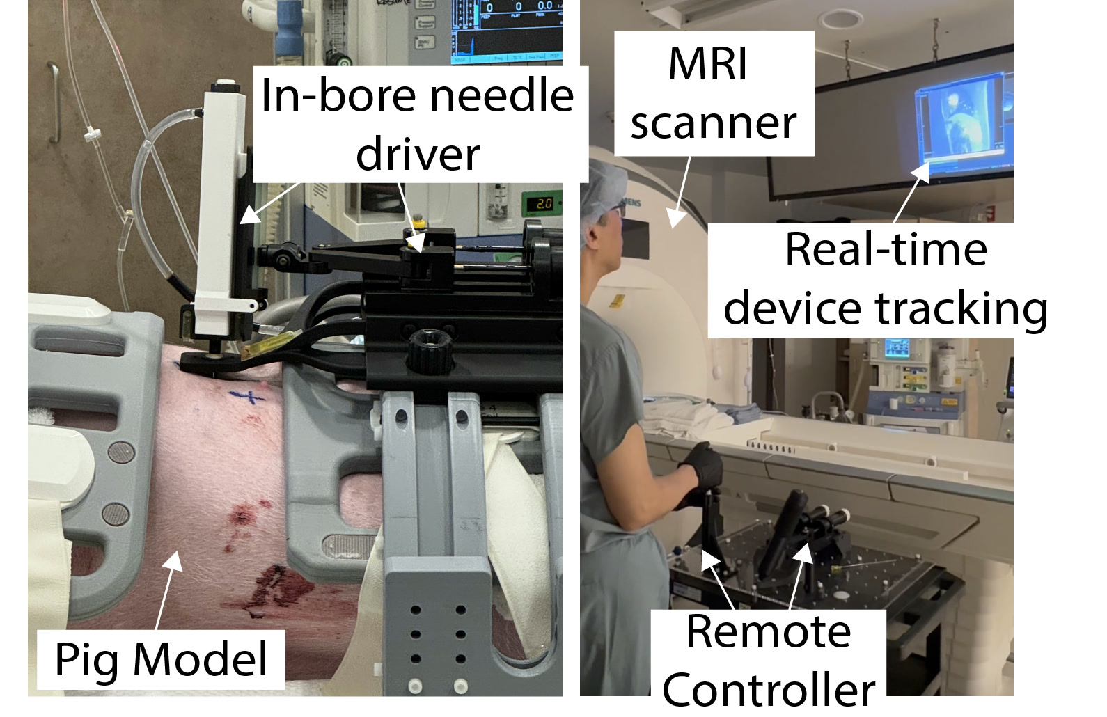
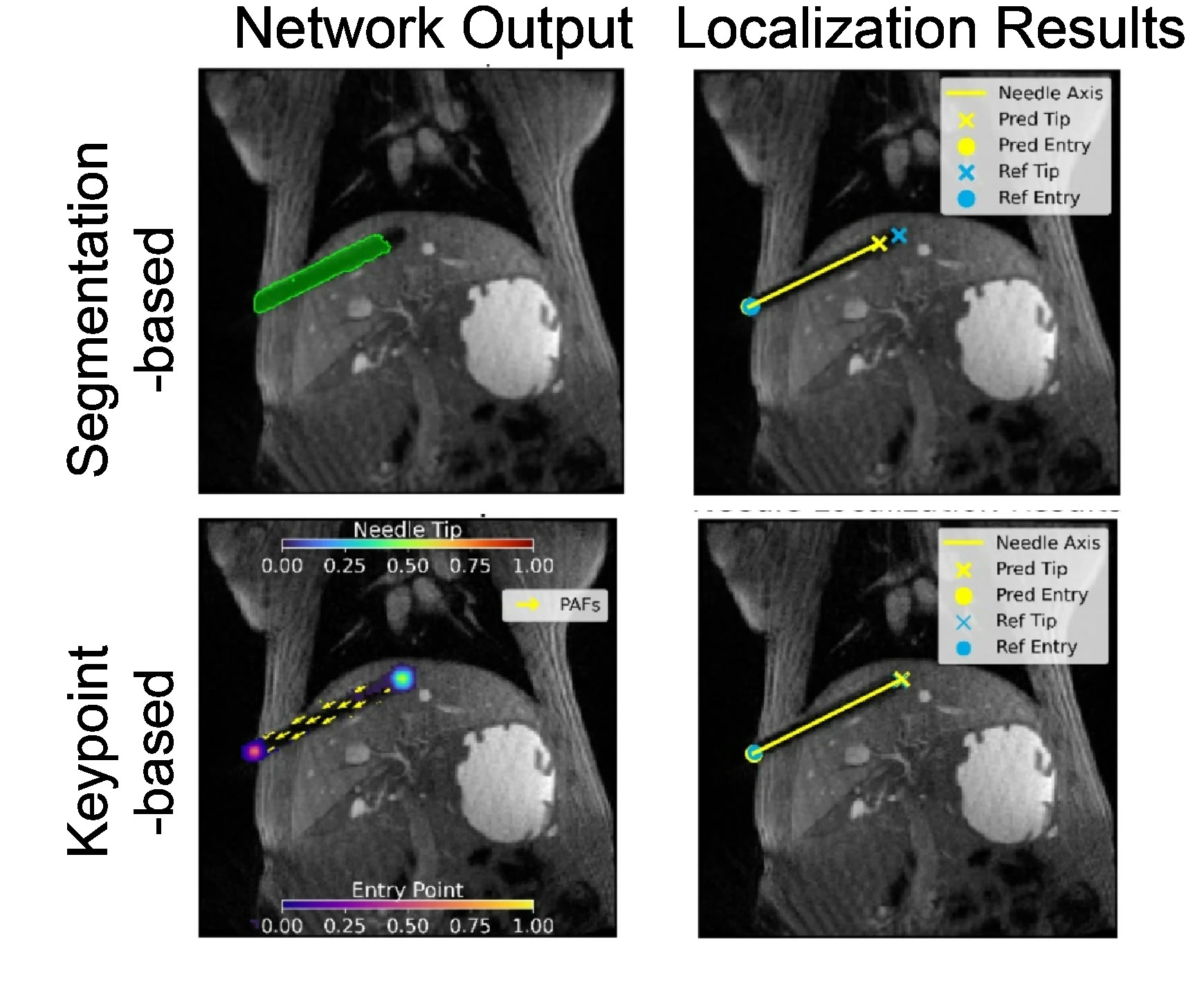
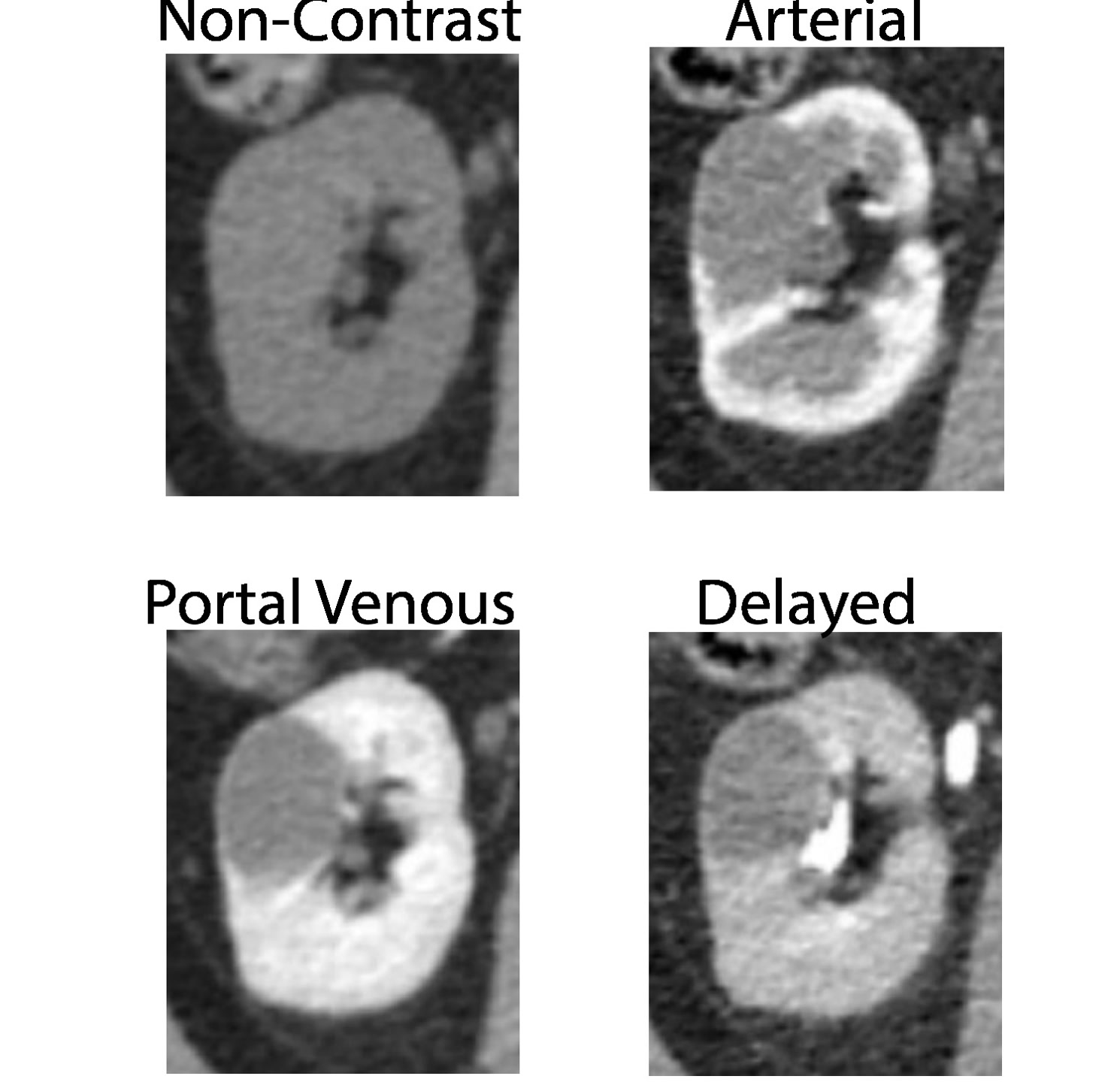
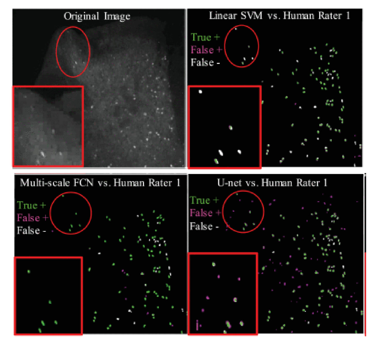
I co-develop deep learning methods for real-time procedural guidance and clinical image analysis. This includes a keypoint detection network for needle localization during MRI-guided interventions that requires minimal annotation (<2 mm error, <35 ms inference; patent pending) [7], 2.5D deep learning frameworks for automated renal tumor grading on the UCSF RMaC dataset (800+ subjects) [8], a U-Net pharmacokinetic estimation framework for hyperpolarized ¹³C MRI [9], and automated neuron segmentation pipelines benchmarking deep learning against classical methods [10].
Machine LearningImage SegmentationDevice TrackingPharmacokinetic ModelingLarge-Scale Clinical DataReal-Time Inference
Pre-clinical translation and validation
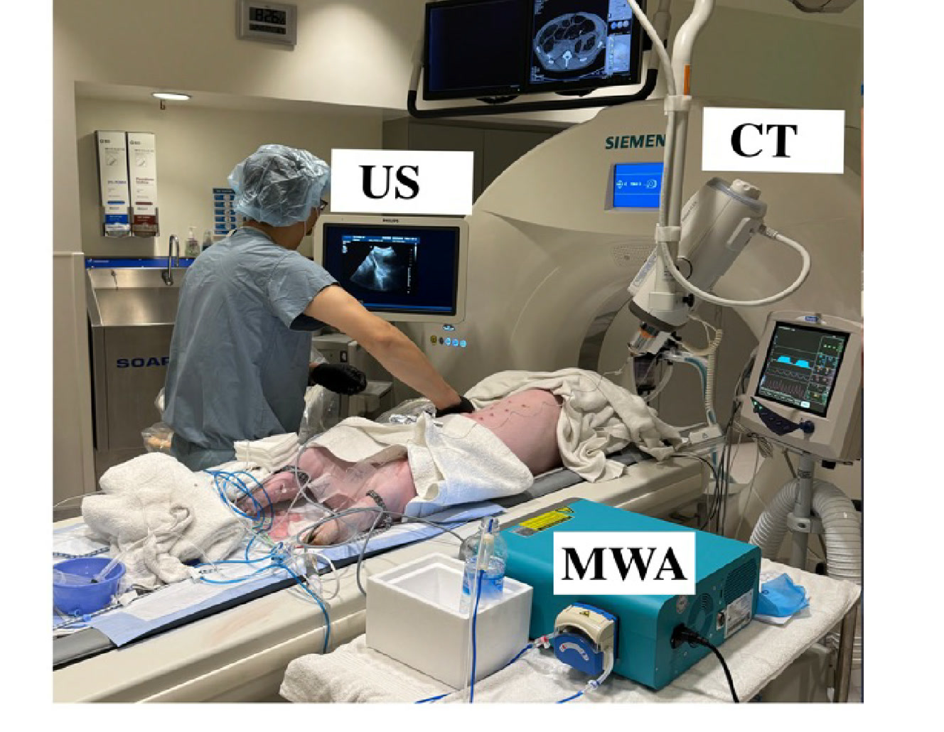
The technical innovations above only matter if they work in practice. Working shoulder-to-shoulder with clinicians, I was a core contributor to UCLA’s pre-clinical MRI-guided intervention program across various imaging platforms (Siemens 3T/0.55T, Bruker 3T), therapeutic modalities (microwave ablation, HIFU, biopsy, surgical robotics), and teams spanning radiology, pharmacology, engineering, and industry — culminating in successful demonstrations from phantom testing through ex vivo validation to in vivo animal models including Oncopig and murine cancer models [4][5]. In collaboration with Dr. David Lu (UCLA Interventional Radiology), the Chiang Lab, and the MAC Lab (UCLA Mechanical & Aerospace Engineering).
MRICTUltrasoundMicrowave AblationHIFUSurgical RoboticsAnimal ModelsRapid PrototypingSystem IntegrationCross-Functional CollaborationPhantom-to-In Vivo Validation